MPI Theory
Magnetic Particle Imaging (MPI) Technology
How does MPI work?
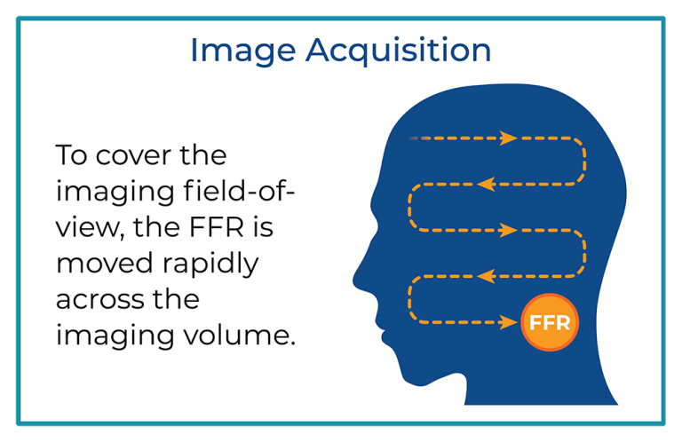
An MPI imager directly detects a tracer known as a Super-Paramagnetic Iron Oxide particle or SPIO. The technology detects only the SPIOs and does not see tissue. These tracers can be modified to suit the application desired. For example, suppose tracers selectively label immunotherapy cells such as CAR T-cells. In that case, we can use MPI to image the distribution of those cells after patient administration. After tracer administration, the patient is positioned in the imaging region of the MPI scanner, and we acquire a three-dimensional image of the tracer distribution.
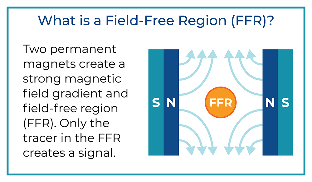
Key to the MPI scanner operation is the strong magnetic field gradient produced inside the imaging volume by the main magnet. Inside this magnetic field gradient, a small region with low magnetic field strength exists, known as the field-free region or FFR. Outside of the FFR, the magnetic field is strong. You can see what this field looks like in the figure, where the magnets are shown in blue/green and the FFR in orange.
MPI or MRI?
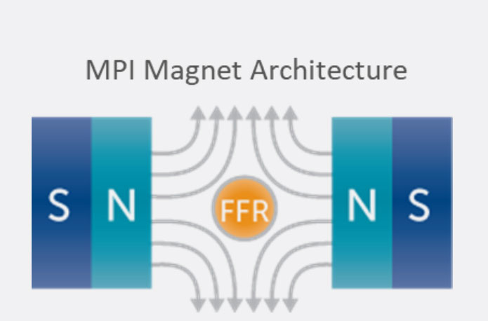
Magnetic Particle Imaging (MPI) and Magnetic Resonance Imaging (MRI) are fundamentally different techniques despite apparent similarities in acronyms and the use of magnets. MRI detects the resonance of nuclear spins (typically the H nucleus) and is generally used for anatomical imaging. MPI detects magnetization in nanoparticles and is used mainly for cellular imaging. These two technologies also use different main magnet architectures.
Two strong magnets pointing at each other produce a magnetic field gradient with an FFR at the center. The FFR is then rapidly moved across the sample to produce an image using strong gradients (T/m) and weak field strengths (mT).
The imaging system’s resolution, sensitivity, and contrast are driven by the interaction between the imager’s main field gradient and the superparamagnetic iron oxide (SPIO) tracer’s magnetic properties. Additionally, the ability of a tracer to visualize an application, such as looking at the immune system, is driven by the nanoparticle’s behavior in tissue.
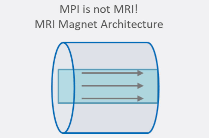
MRI creates a uniform field to produce an image using weak gradients (mT/m) and strong field strengths (T).